Electrocochleography (ECoG) is a specialized electrophysiological test now available in Dhaka at Vertigo & Balance Center, used to detect fluid pressure changes deep inside the inner ear. It’s one of the most valuable tools for confirming or ruling out Ménière’s disease — especially when symptoms like fluctuating hearing loss, ear fullness, and episodic vertigo make the diagnosis unclear. Here’s what an ECoG test in Dhaka involves and what your results mean.

MINUTES
ELECTRODE OPTIONS
Stimuli LOW-RISK
Electrocochleography (ECoG) is an electrophysiological test used to measure the electrical potentials generated by the cochlea and the auditory (eighth cranial) nerve in response to sound stimulation.
The test records tiny electrical signals arising from the inner ear and the auditory nerve pathway using an electrode placed close to the cochlea. ECoG is especially valuable in the evaluation of endolymphatic hydrops and Ménière’s disease, as well as in intraoperative monitoring.
ECoG provides objective electrophysiological data that supports and refines the clinical diagnosis of inner ear fluid pressure disorders.
Sound stimulation causes the cochlear hair cells to generate receptor potentials, and the auditory nerve fibers generate an action potential in response. ECoG records three principal components:
In endolymphatic hydrops, distension of the endolymphatic compartment alters the mechanics of the basilar membrane, typically increasing the amplitude of the summating potential relative to the action potential (an elevated SP/AP ratio).
The patient is seated comfortably in a quiet test environment
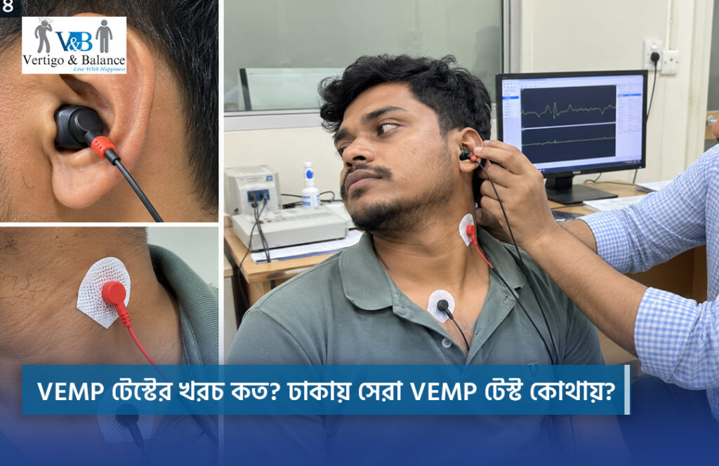
An electrode is placed on the tympanic membrane, ear canal, or transtympanically, according to protocol
Click or tone-burst stimuli are presented through an insert earphone
Electrical responses are recorded, averaged, and analyzed
ECoG is a low-risk test. Extratympanic recording is non-invasive and generally well tolerated. Transtympanic (needle) recording involves brief mild discomfort at the time of electrode placement. All procedures are performed under professional supervision, and the test can be paused if needed.
ECoG results provide objective electrophysiological information regarding cochlear and auditory nerve function, particularly in the assessment of endolymphatic hydrops. Interpretation should be performed by trained clinicians and correlated with patient history, audiometric findings, and additional vestibular investigations.
Clinical Use: Diagnostic Evaluation and Treatment Planning
Tympanic membrane (TM) electrode; ear canal (extratympanic) electrode; transtympanic (needle) electrode — used when a stronger, more precise signal is required. Extratympanic recording is non-invasive and more commonly used in outpatient clinical practice, while transtympanic recording provides a stronger signal and is often reserved for select diagnostic or surgical cases.
Click stimuli — broadband, used for general cochlear and nerve response. Tone-burst stimuli — frequency-specific responses. Stimuli are typically delivered through insert earphones at controlled intensity levels while responses are averaged to improve the signal-to-noise ratio.
Measurement of SP and AP amplitudes; calculation of the SP/AP amplitude ratio; comparison between ears when bilateral testing is performed.
• Requires patient cooperation and a quiet test setting
• Transtympanic recording is more invasive than extratympanic recording
• Signal quality can be affected by electrode placement and impedance
• Findings must be interpreted alongside audiometric and vestibular test results
ECoG should be combined with other audiological and vestibular tests for comprehensive evaluation.
Normal ECoG: SP/AP ratio within the normal reference range and well-defined AP waveform morphology.
Abnormal ECoG: elevated SP/AP ratio, widened AP waveform, or reduced amplitude responses.
Findings may indicate:
Electrocochleography (ECoG) is a specialized electrophysiological test that measures electrical potentials in the inner ear (cochlea) and auditory nerve in response to sound. It is primarily performed to detect abnormal fluid pressure (endolymphatic hydrops) and confirm conditions like Ménière’s disease.
Yes, ECoG is one of the most effective objective diagnostic tools for Ménière’s disease. By evaluating the SP/AP ratio (Summating Potential to Action Potential ratio), ECoG identifies inner ear fluid pressure changes, helping specialists confirm Ménière’s disease even when standard hearing tests or MRIs are inconclusive.
A standard audiogram only measures how loudly or clearly you hear sound frequencies. In contrast, an ECoG test records the exact electrical signals generated by inner ear hair cells and the hearing nerve, directly detecting fluid pressure disorders that regular hearing tests cannot show.
Vestibular migraines and Ménière’s disease share very similar symptoms, such as episodic vertigo and ear fullness. ECoG helps differentiate them by measuring inner ear pressure: an elevated SP/AP ratio typically points to Ménière’s disease, whereas a normal ECoG result combined with migraine symptoms suggests a vestibular migraine.
Non-invasive extratympanic ECoG (where gentle surface electrodes are placed on the eardrum or ear canal) is completely painless and well-tolerated. If transtympanic recording is needed, it involves minimal, momentary discomfort during electrode placement under professional clinical supervision.
Vestibular assessments performed
Dedicated diagnostic test modalities
BMDC-registered vestibular specialists
Established-Concept Tower, Panthapath
Vestibular Specialist & Neurotologist BMDC Reg. 65718-Founder & Chief Consultant
Vestibular Rehabilitation Specialist BMDC Reg. A-105382-AIB-Certified, VRT Center Lead
Trained Vestibular Professional
Concept Tower (3rd Floor), Room: 337 & 338. 68-69, Green Road, Dhaka-1205